Metamorphosia tests by Metrovision
- Home
- Visual function tests
- Vision psychophysics
- Metamorphopsia
(This feature is not available for sale in the United States of America)
Metamorphopsia and ARMD
The early detection and intervention are important to prevent or delay vision loss in age related macular degeneration (ARMD).
Retinal metamorphopsia occur in the early stage of ARMD as a result of elevations of the pigment epithelium. These metamorphopsia are usually evaluated with the Amsler chart. However, this test suffers from several limitations including the difficulty of self reporting, the absence of fixation monitoring, the absence of quantification and problems with filling-in phenomenon.

Methodology
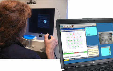
This program provides an automated detection of metamorphopsia and the follow-up of their evolution. The test is similar to a perimetry test. Local patterned stimuli are presented in a pseudo random sequence and the patient task is to indicate whether the pattern is distorted or not. The test duration is about 150 seconds including controls for attention and fixation errors.
This program is available on the Moncv3 and MonPackONE systems
These systems include a built-in fixation monitoring device.
A standard PC operating under Windows is used for the control of the instrument as well as for the storage and printout of results.
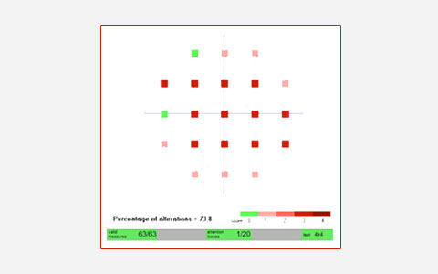
Quantitative analysis
The program provides a standardized report including a map of probability of altered locations and the total percentage of locations with alterations.
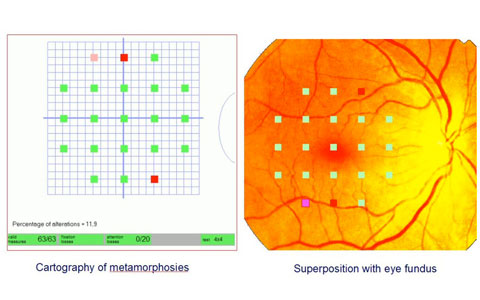
Comparison with the eye fundus
The program allows the comparison of the metamorphopsia with the eye fundus.
The image of the eye fundus is imported as an image file either through the computer network
or by USB key, CDROM etc. The operator identifies by a simple click the position of the fovea
and papilla and the program automatically performs the scaling and repositioning the eye fundus image.
Bibliography and references
PDF âComparative Evaluation of Visual Outcomes in Combined Cataract and Vitrectomy for Idiopathic Epiretinal Membrane with an Advanced or Conventional Intraocular Lens
PDF âAdditional measures of macular function beyond visual acuity
PDF âCorrelations among metamorphopsia test scores, optical coherence tomography findings and multifocal electroretinogram responses in epiretinal membrane patients
PDF âCorrelation between Quantification of Metamorphopsia and Optical Coherence Tomography Findings in Patients with Epiretinal Membrane
PDF âEvaluation des mÃĐtamorphopsies