Test of macular pigment by Metrovision
- Home
- Visual function tests
- Vision psychophysics
- Macular pigment
(This feature is not available for sale in the United States of America)
Macular pigment and ARMD
The purpose of this test is to detect subjects with a low density of macular pigment.
The macular pigment is present in the central part of the retina. It has an antioxidant effect
due to a high concentration of hydroxy-carotenoids (luthein and zeaxanthin).
It plays a protective role against the development of age related macular degeneration (Haddad et al, 2006, Whitehead et al, 2006).
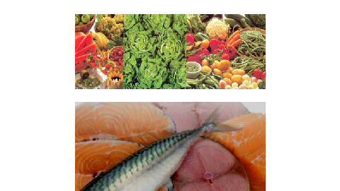
Furthermore, it has been shown that the density of macular pigments is highly related to nutrition (Crochet et al, 2009)
and that a change in diet or the use of nutritional supplements modifies the macular pigment density (Hammond et al, 1997).
Methodology
It has been shown that the macular pigment absorbs blue light.
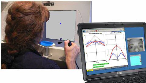
This is why the program evaluates the density of the macular pigment by comparing the thresholds
of perception of blue light and red light with a staircase technique similar to the technique used in automated perimetry.
This technique presents several advantages:
• the staircase thresholding technique is much easier for patients than the heterochromatic flicker technique.
• it does not require the dilation of pupils
• it is quite fast (less than 3 minutes per eye).
This program is available on the Moncv3 and MonPack systems.
These systems include a built-in fixation monitoring. A standard PC operating under Windows is used for the control of the instrument as well as for the storage and printout of results.
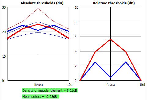
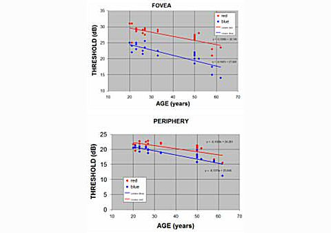
Results from normal subjects
The curves hereby show results from normal subjects for the detection thresholds of blue and red light
measured at the fovea and in pericentral locations.
In normal subjects, the blue thresholds present a relative attenuation of about 0.6 log units in the
fovea location, indicating the presence of macular pigment that absorbs blue light.
Analysis of the results
The first step of the analysis is to compensate for the alteration of blue thresholds due to intraocular media transparency.
This is achieved by substracting the measurement at the pericentral location (affected only by intraocular media)
from the measurement at the fovea (involving both pigment and intraocular media).
In a second step, the result is compared to an age corrected normal data base that has been developped from a population
of subjects with normal eyes and a nutritionaly recommended diet (more details in the publication cited in references).