Metrovision's Visual field tests
- Home
- Visual function tests
- Vision psychophysics
- Visual field tests
Introduction
Several solutions are proposed for the assessment of the visual field.
They correspond to specific clinical applications:
• Standard automated perimetry for the detection and follow-up of glaucoma
• Goldmann perimetry for neuro-ophthalmology and difficult patients
• Attraction perimetry for young children and handicapped subjects
• Micro-perimetry for the comparison with the eye fundus or OCT
Standard automated perimetry (SAP)
The standard static perimetry procedures include full threshold as well as fast threshold algorithms.
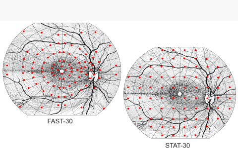
Two series of test grids are proposed :
The STAT tests use the conventional grids of test points, with a constant spacing between points.
The FAST tests (Fiber Adapted Static Testing Perimetry) use a distribution of points optimized
as a function of the most frequent alterations of the retina and optic nerve.
Test points chosen on the basis of physiology and pathology provide more useful information in less time.
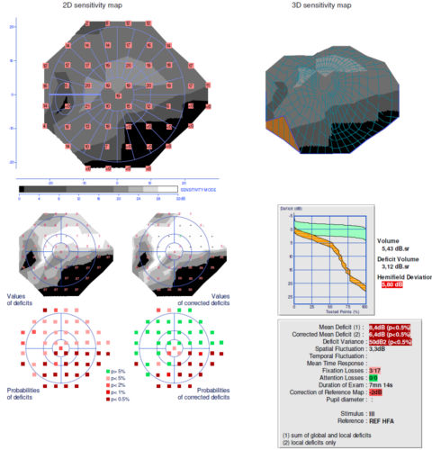
Standard automated perimetry report
Modern graphic technology facilitates the interpretation of the visual field
The standard report for SAP includes:
• a modern graphic display combining the position and values of measurements with a true grey scale map
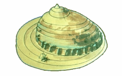
• a 3D representation for a better understanding of the topography of the field
• a statistical analysis with comparison with normative data
• global indexes (mean defect, ..) as well as information about the reliability of test (fixation errors, attention errors)
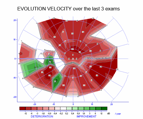
Follow-up analysis
The follow-up program generates a list of all perimetry exams performed on the same eye of the same patient.
It shows the evolution of the global indexes over time (mean deficit and corrected mean deficit).
It also displays the evolution velocity map indicating with a color code the rate of evolution of the different locations of the field.
The evolution velocity map allows to distinguish between evolution due to glaucoma, cataract and ARMD processes
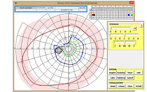
Goldmann perimetry
Goldmann perimetry is still useful in the 21st century
Goldmann perimetry still has several important clinical applications:
• for the patients who cannot reliably perform automated perimetry .
• for severely altered visual fields, it is much faster and efficient than SAP
• for the confirmation of deficits found in automated perimetry
Goldmann perimetry is entirely controled "manually" with the computer mouse.
The operator can select the Goldmann stimulus parameters, control the stimulation presentation and draw the isopters.
Goldmann perimetry is available on all Metrovision's visual field instruments: MonCvONE (for full field), MonCv3 and MonPackONE (for central field).
Attraction perimetry
Attraction perimetry allows testing the visual field in young children
This technique uses the eye movement response of the child. The operator controls the presentation of visual stimuli in the periphery and uses the high quality video to detect the orientation response of the child.
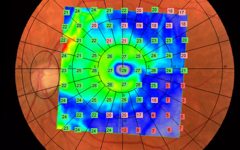
Micro-perimetry
Micro perimetry allows the comparison of the visual field with the eye fundus or OCT
Just download the eye fundus or OCT image before starting the exam.
Click on the fovea and papilla positions for calibation... and the visual field exam
can be realized with the eye fundus image on the background.